
Cardiac Ultrasound at Evendale-Blue Ash Pet Hospital
An echocardiogram, also known as a cardiac ultrasound, is a specialized test used to examine the heart. It’s similar to what humans get when they have a "heart echo."
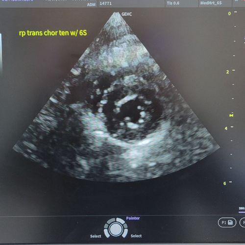
At EBPAH, Dr. Goodman is certified to conduct cardiac ultrasounds on dogs and cats. The test uses sound waves to create pictures of the heart as it beats, showing us how well the heart is working. This test is recommended when we hear a heart murmur or arrhythmia (abnormal rhythm) during your furbaby's examination.
How It Works
When your pet comes in for a cardiac ultrasound, these are the steps that Dr. Goodman and her team will take.
- We clip a small area of fur over the heart.
- We then apply a gel to the chest, typically around the area where the heart is located.
- Next, we place a small device called a "transducer" on the skin.
- This transducer sends out high-frequency sound waves that bounce off the heart and return to the machine.
- The ultrasound machine turns these sound waves into moving images of the heart, which helps Dr. Goodman see things like:
- The size of the heart
- The thickness of the heart walls
- How well the heart is pumping blood
- If there are any problems with the heart valves.
The Benefits of Cardiac Ultrasound
Cardiac ultrasound is really helpful because it can spot issues like heart disease, heart murmurs, or abnormal blood flow, without needing to do surgery or use radiation. It’s painless, non-invasive, and provides real-time images, which can help the team at Evendale-Blue Ash Pet Hospital make informed decisions about the best way to treat your furbaby.
In short, a cardiac ultrasound provides a detailed, live view of your pet's heart, allowing us to identify any potential issues early and ensure your pet remains healthy.