
Cardiac Ultrasound for Cats
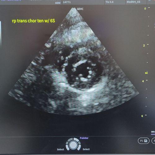
An echocardiogram, or cardiac ultrasound, is a specialized test used to evaluate the heart’s structure and function. It’s similar to what humans receive when they undergo a “heart echo.”
At EBPAH, Dr. Goodman is certified to perform cardiac ultrasounds on cats. The procedure uses sound waves to create moving images of the heart, allowing us to see how well it’s working. This test is often recommended when we detect a heart murmur or an irregular rhythm during your cat’s examination.
It provides a real-time view of the heart, including:
- Heart chambers and valves
- Blood flow patterns
- Heart wall motion
- Pumping efficiency
How does an echocardiogram help diagnose heart conditions in cats?
An echocardiogram offers a detailed, live view of the heart, making it one of the most valuable tools for diagnosing feline heart disease. It allows veterinarians to detect changes in structure, movement, and blood flow that other diagnostic methods can’t reveal.
Here’s what an echocardiogram can help us determine:
- Identifies Structural Changes such as enlarged heart chambers or thickened heart walls.
- Evaluates Valve Health for regurgitation (leakiness) or narrowing that affects blood flow.
- Measures Pumping Ability by assessing contraction and relaxation phases.
- Shows Blood Flow with Doppler imaging to evaluate speed and direction.
- Tracks Progress or Response to Treatment over time.
This non-invasive, painless procedure provides critical information that can’t be obtained from X-rays or a stethoscope alone.
What’s the difference between an echocardiogram and an electrocardiogram for cats?
An echocardiogram shows what the heart looks like and how it moves. An electrocardiogram (ECG or EKG) records the electrical activity of the heart, displaying rhythm, rate, and irregularities in the heartbeat.
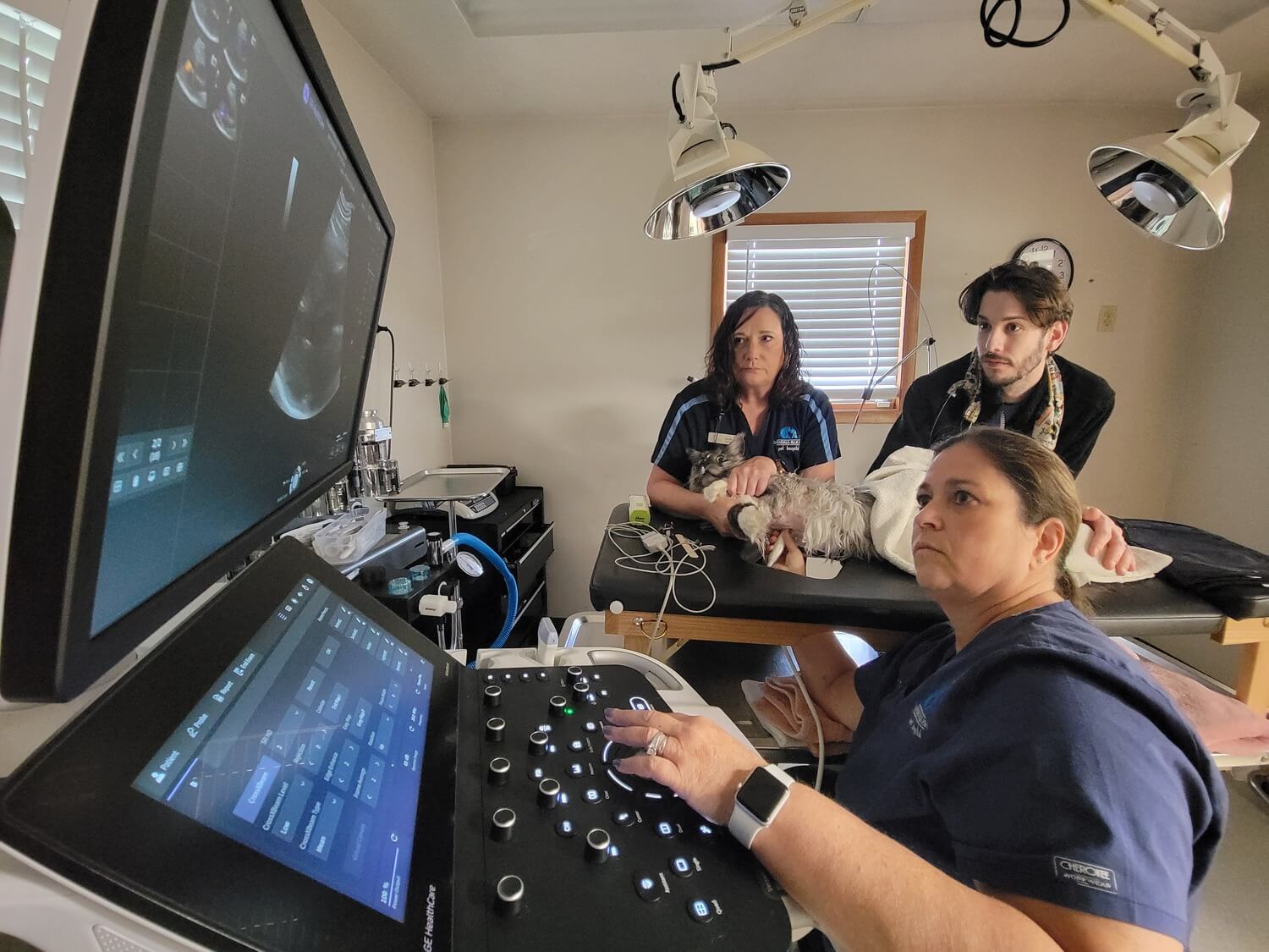
What to Expect When Your Cat Needs an Echocardiogram

When your cat comes in for a cardiac ultrasound, Dr. Goodman and her team will follow these steps:
The Cardiac Ultrasound Procedure for Cats:
- A small patch of fur is clipped over the heart area.
- Ultrasound gel is applied to the chest to improve sound wave transmission.
- A handheld device called a “transducer” is placed against the skin.
- The transducer sends sound waves into the chest, which bounce back from the heart structures.
- The machine converts these echoes into live images of the heart in motion.
When We Recommend an Echocardiogram for Cats
- To evaluate a heart murmur
- To confirm or monitor heart disease (e.g., hypertrophic cardiomyopathy)
- To investigate congenital heart defects (especially in kittens)
- To assess fluid buildup around the heart (pericardial effusion)
- To evaluate overall heart size and function
How is a heart murmur evaluated using an echocardiogram?
In cats, a heart murmur can be an early sign of heart disease. An echocardiogram allows us to see the cause—often related to abnormal blood flow—and to determine the severity of the problem.
Is an echocardiogram safe and comfortable for cats?
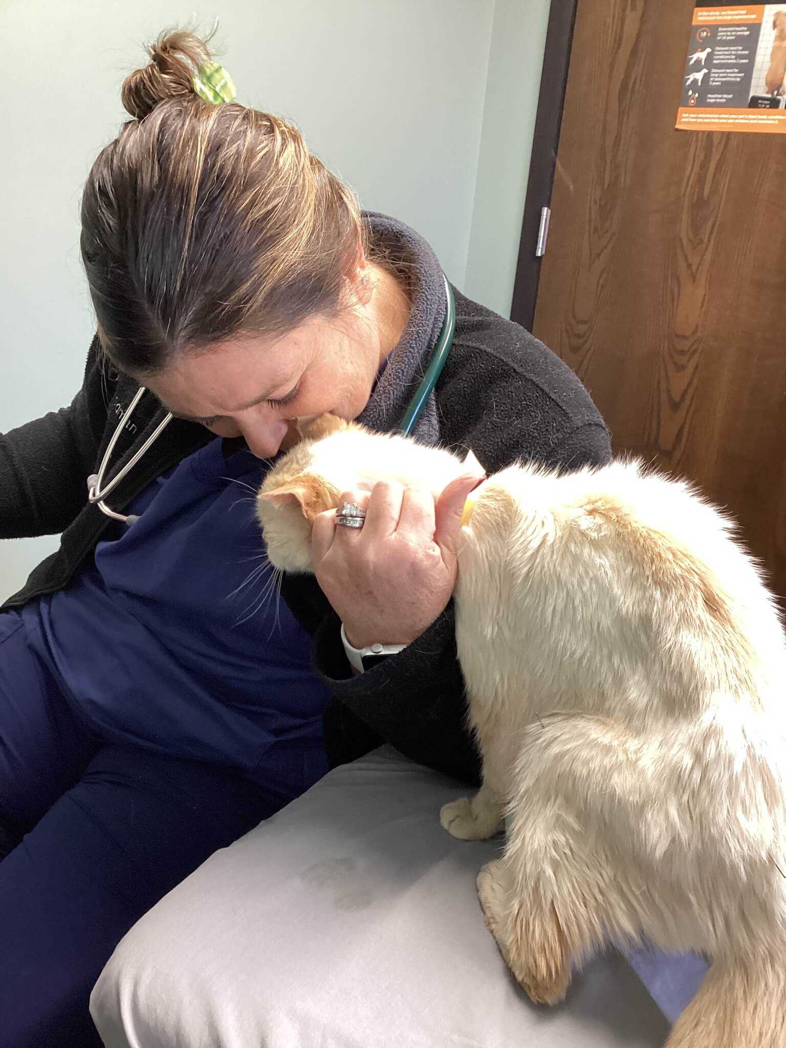
Yes. It is considered very safe.
- It’s non-invasive and uses sound waves, not radiation.
- There are no known risks associated with the procedure.
- Most cats tolerate it well while calmly lying on their side or chest.
- Only a small patch of fur is shaved to improve imaging.
- Ultrasound gel and a probe are used for clear images.
If your cat is showing signs of heart disease or you’d like to learn more about our feline cardiology services, schedule an appointment with Dr. Goodman to discuss cardiac ultrasound testing.