
Cardiac Ultrasound for Dogs
An echocardiogram, also known as a cardiac ultrasound, is a specialized test used to examine the heart. It’s similar to what humans get when they have a "heart echo."
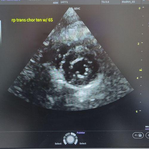
At EBPAH, Dr. Goodman is certified to conduct cardiac ultrasounds on dogs. The test uses sound waves to create pictures of the heart as it beats, showing us how well the heart is working. This test is recommended when we hear a heart murmur or arrhythmia (abnormal rhythm) during your furbaby's examination.
It shows the heart's structure and function in real time, including:
- Heart chambers and valves
- Blood flow through the heart
- Heart wall motion
- How well the heart is pumping
How does an echocardiogram help diagnose heart conditions in dogs?
An echocardiogram helps diagnose heart conditions in dogs by providing a detailed, real-time view of the heart’s structure and function. It’s one of the most important tools veterinarians use to evaluate canine and feline heart disease.
Here's what an echocardiogram can do for your pet:
- Identifies Structural Abnormalities, such as enlarged heart chambers and thickened heart walls.
- Evaluates Valve Function for valve regurgitation (or “leaky” valves) and narrowing of the valves that may restrict blood flow.
- Measures Heart Pumping Efficiency by assessing how well the heart contracts and relaxes.
- Visualizes Blood Flow using Doppler imaging to show direction and speed of blood flow.
- Monitors Disease Progression or Treatment Response by tracking changes over time.
An echocardiogram is non-invasive, painless, and provides crucial information that can’t be gained through X-rays or listening to the heart alone.
What’s the difference between an echocardiogram and an electrocardiogram for dogs?
An echo shows what the heart looks like and how it moves. An electrocardiogram (ECG or EKG) shows how the heart's electrical system is functioning. An ECG shows the heart rhythm and rate, irregular heartbeats (arrhythmias), and electrical conduction issues.
What to Expect When Your Pet Needs an Echocardiogram
When your pet comes in for a cardiac ultrasound, these are the steps that Dr. Goodman and her team will take.
The Cardiac Ultrasound Procedure for Dogs:
- We clip a small area of fur over the heart.
- We then apply a gel to the chest, typically around the area where the heart is located.
- Next, we place a small device called a "transducer" on the skin.
- This transducer sends out high-frequency sound waves that bounce off the heart and return to the machine.
- The ultrasound machine turns these sound waves into moving images of the heart.
Why An Echocardiogram Would Be Recommended for Your Pet
- To evaluate heart murmurs
- To confirm or monitor heart disease (e.g., mitral valve disease, cardiomyopathy)
- To check for congenital defects (especially in puppies)
- To assess fluid around the heart (pericardial effusion)
- To evaluate heart size and function
How is a heart murmur evaluated using an echocardiogram?
A heart murmur in a dog is often the first clue that something may be wrong with the heart, and an echocardiogram is the gold standard for evaluating it. A murmur is usually due to abnormal blood flow, and the echocardiogram can determine exactly why.
Is an echocardiogram safe and comfortable for dogs?
Yes, it is safe!
- It's non-invasive and uses ultrasound, not radiation.
- There are no known risks associated with the procedure.
- Most dogs tolerate the procedure well while lying calmly on their side.
- A small area of fur is typically shaved on the chest to allow for clear imaging.
- Ultrasound gel is applied to help transmit sound waves, and a handheld probe is used.
If your dog is showing signs of a heart condition or you have questions about our veterinary cardiology services, schedule an appointment with Dr. Goodman to learn more about dog cardiac ultrasounds.